42 protein synthesis diagram worksheet
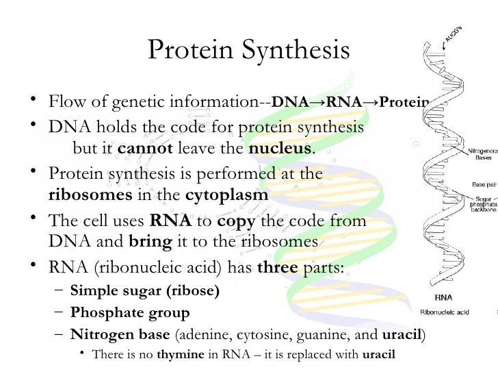
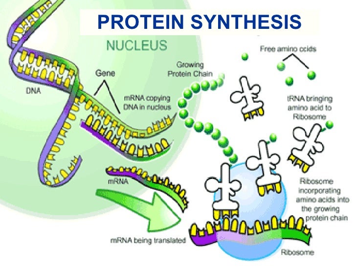
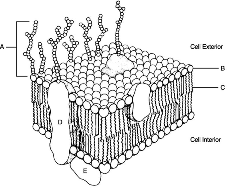
Plant Cell- Definition, Structure, Parts, Functions, Labeled ... Feb 16, 2022 · Figure: Diagram of the cell (plasma) membrane. Source: Wikipedia Structure of the plant cell (plasma) membrane. This is a bilipid membrane that is made up of protein subunits and carbohydrates, with a characteristic semi permeability factor. It surrounds the cell cytoplasm, thus enclosing its content. Functions of the plant cell (plasma) membrane Transcription and Translation | Basic Biology Aug 31, 2020 · During translation, the RNA molecule created in the transcription process delivers information from the DNA to the protein-building machines. DNA → RNA → Protein. DNA and RNA are similar molecules and are both built from smaller molecules called nucleotides. Proteins are made from a sequence of amino acids rather than nucleotides.
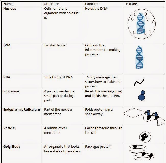
The Structure of Prokaryote and Eukaryote Cells - CliffsNotes The rough ER is the site of protein synthesis in a cell because it contains ribosomes; however, the smooth ER lacks ribosomes and is responsible for producing lipids. Within the ribosomes, amino acids are actually bound together to form proteins. Cisternae are spaces within the folds of the ER membranes.

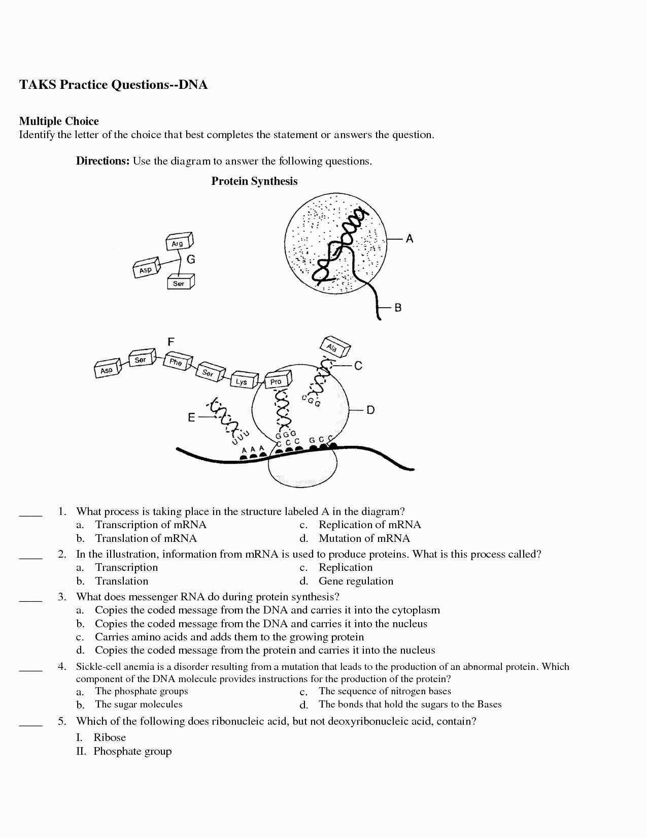
Protein synthesis diagram worksheet
All Biology Study Notes (Alphabetical Order) Protein Databases- Types and Importance; Protein Synthesis Inhibitors- Definition, Examples, Inhibition, Resistance; Protocol of chloroplast isolation; Protocol: Phenol-chloroform extraction of prokaryotic DNA; Protocooperation Interaction- Definition and Examples; Protozoa- Definition, Characteristics, Classification, Examples Animal Cell Anatomy & Diagram - Enchanted Learning Spherical body containing many organelles, including the nucleolus. The nucleus controls many of the functions of the cell (by controlling protein synthesis) and contains DNA (in chromosomes). The nucleus is surrounded by the nuclear membrane. Ribosome Small organelles composed of RNA-rich cytoplasmic granules that are sites of protein synthesis. The Cell Cycle - CELLS alive Interphase generally lasts at least 12 to 24 hours in mammalian tissue. During this period, the cell is constantly synthesizing RNA, producing protein and growing in size. By studying molecular events in cells, scientists have determined that interphase can be divided into 4 steps: Gap 0 (G0), Gap 1 (G1), S (synthesis) phase, Gap 2 (G2).
Protein synthesis diagram worksheet. Biology corner transcription and translation answer key caac gki ecdc aa iahe ksb jah cb bf aaaa oocb gc gih or grpl ba ejfb fg amh ba idbp lk caj ai bdb gd ichj deh gafj fg bbba The Cell Cycle - CELLS alive Interphase generally lasts at least 12 to 24 hours in mammalian tissue. During this period, the cell is constantly synthesizing RNA, producing protein and growing in size. By studying molecular events in cells, scientists have determined that interphase can be divided into 4 steps: Gap 0 (G0), Gap 1 (G1), S (synthesis) phase, Gap 2 (G2). Animal Cell Anatomy & Diagram - Enchanted Learning Spherical body containing many organelles, including the nucleolus. The nucleus controls many of the functions of the cell (by controlling protein synthesis) and contains DNA (in chromosomes). The nucleus is surrounded by the nuclear membrane. Ribosome Small organelles composed of RNA-rich cytoplasmic granules that are sites of protein synthesis. All Biology Study Notes (Alphabetical Order) Protein Databases- Types and Importance; Protein Synthesis Inhibitors- Definition, Examples, Inhibition, Resistance; Protocol of chloroplast isolation; Protocol: Phenol-chloroform extraction of prokaryotic DNA; Protocooperation Interaction- Definition and Examples; Protozoa- Definition, Characteristics, Classification, Examples

Protein Synthesis Diagram Worksheet - Promotiontablecovers

Pinterest • The world’s catalog of ideas

Building Dna Gizmo Worksheet Answers / Rna Protein Synthesisse - Powell ...

Protein Synthesis Diagram Worksheet Answer Key - Aflam-Neeeak
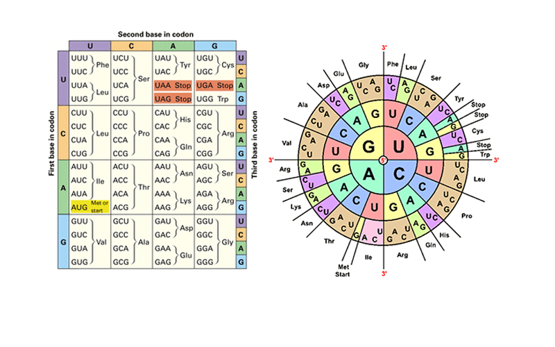
Amino Acid Codon Chart Circle - The Chart
Cell Cycle, Dna, And Protein Synthesis Notes New
Seventh grade Lesson Cells: Protein Production Part 1
Protein Synthesis Worksheet – Wiring Diagram — db-excel.com

Media Portfolio

Wiring And Diagram: Diagram Protein Synthesis Worksheet
Protein Synthesis

3 Protein Synthesis Worksheet Answers | FabTemplatez
Print Chapter 3 Cells: The Living Units flashcards | Easy Notecards

Protein Synthesis Diagram Worksheets

Protein Synthesis Diagram Analysis Worksheet Answer Key - Aflam-Neeeak
0 Response to "42 protein synthesis diagram worksheet"
Post a Comment